
Del i serien Advanced Chemical Products and Materials
Fluorescent Dyes
Synthesis and Biomedical Applications
Inbunden, Engelska, 2026
2 199 kr
Beställningsvara. Skickas inom 11-20 vardagar. Fri frakt för medlemmar vid köp för minst 249 kr.
Provides comprehensive and up-to-date knowledge on the fluorescence technology in biomedical applications.
Produktinformation
- Utgivningsdatum2026-11-18
- Mått170 x 244 x undefined mm
- FormatInbunden
- SpråkEngelska
- SerieAdvanced Chemical Products and Materials
- Antal sidor416
- FörlagWiley-VCH Verlag GmbH
- ISBN9783527353460
Tillhör följande kategorier
Hoppa över listan


Du kanske också är intresserad av
- Nyhet
- Nyhet
Genesis, Volume 2: Creation, Fall, and Redemption, (Chapters 20-50), a 13-Lesson Study
Zachary Groff
Häftad
249 kr
Affirmations for Artists: A Complete Guide to Building a Positive Creative Mindset
Phd Eric Maisel
Häftad
319 kr
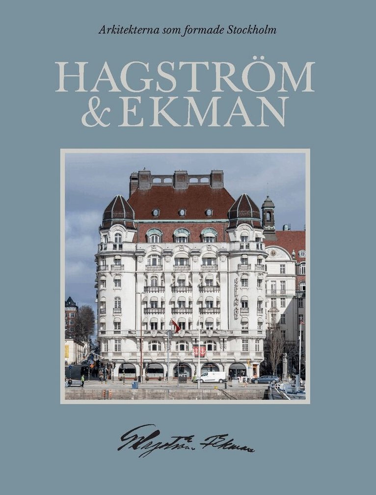
Arkitekterna som formade Stockholm: Hagström & Ekman
Sandra Nolgren, Johan Mårtelius, Martin Rörby, Tove Grönroos, Marie Ekblad, Gustav Bergström, Mikael Traung, Ann Katrin Pihl Atmer
Inbunden
549 kr