
Small Animal Gastroenterology
Inbunden, Engelska, 2008
1 579 kr
Beställningsvara. Skickas inom 5-8 vardagar. Fri frakt för medlemmar vid köp för minst 249 kr.
Written by authors from around the world with a variety of backgrounds using evidence-based medicine, this book has two main sections. The first covers the diagnosis of gastrointestinal disorders. This section contains chapters on various different diagnostic methods and on the most commonly observed clinical signs of gastrointestinal disease presented in dogs and cats, acute gastrointestinal signs, chronic vomiting, chronic diarrhea, and weight loss.The second section focuses on specific diseases of the gastrointestinal tract. Each component of the GI tract is discussed separately with another section focusing on diseases that affect more than one organ of the gastrointestinal tract. The book is well referenced and fully illustrated throughout. It is concise but detailed enough for both veterinarians in training and in practice.
Produktinformation
- Utgivningsdatum2008-04-30
- Mått210 x 276 x 25 mm
- Vikt1 500 g
- FormatInbunden
- SpråkEngelska
- Antal sidor384
- Upplaga1
- FörlagSchlutersche
- ISBN9783899930276
Tillhör följande kategorier
- AuthorsAbbreviationsPrefacePart I Diagnosis ofGastrointestinal Disorders1 Diagnostic Tools1.1 Clinical History1.1.1 Introduction1.1.2 History of specific gastrointestinal signs1.1.2.1 Dysphagia and regurgitation1.1.2.2 Gagging1.1.2.3 Vomiting1.1.2.4 Retching1.1.2.5 Diarrhea1.1.2.6 Other stool abnormalities1.1.2.7 Flatulence and borborygmus1.1.2.8 Dyschezia1.1.2.9 Constipation1.1.2.10 Fecal incontinence1.1.2.11 Anal pruritus1.1.2.12 Abdominal pain1.1.3 Dietary history1.2 Physical Examination1.2.1 Introduction1.2.2 General physical examination1.2.2.1 Skeletal growth and development1.2.2.2 Body condition1.2.2.3 Mental status1.2.2.4 Abnormalities in posture and locomotion1.2.2.5 Mucous membranes1.2.2.6 Peripheral lymph nodes1.2.2.7 Skin and subcutaneous tissue1.2.2.8 Body temperature1.2.2.9 Pulse rate1.2.2.10 Respiratory rate1.2.3 Examination of the gastrointestinal tract1.3 Diagnostic Imaging1.3.1 Introduction1.3.2 Oropharynx1.3.2.1 Structural abnormalities1.3.2.2 Functional Disorders1.3.3 Esophagus1.3.3.1 Generalized esophageal dilation1.3.3.2 Segmental esophageal dilation1.3.4 Stomach1.3.4.1 Gastric dilation and volvulus1.3.4.2 Gastric causes of chronic vomiting1.3.4.3 Diagnosis of delayed gastric emptying1.3.5 Small intestine1.3.5.1 Ileus1.3.5.2 Partial obstructions1.3.5.3 Complete obstructions1.3.5.4 Functional ileus1.3.5.5 Detecting ileus with ultrasound1.3.5.6 Complicated ileus1.3.5.7 Chronic diarrhea1.3.5.8 Diffuse bowel wall infiltration1.3.5.9 Gastrointestinal hemodynamic assessmentwith Doppler ultrasound1.3.6 Large intestine1.3.7 Liver and biliary tract1.3.7.1 Hepatic parenchymal disease1.3.7.2 Non-obstructive biliary tract disease1.3.7.3 Obstructive disease1.3.7.4 Interventional procedures of the liverand biliary system1.3.8 Pancreas1.3.8.1 Pancreatitis1.3.8.2 Pancreatic neoplasia1.4 Laboratory Tests1.4.1 Laboratory assessment of gastric disease1.4.1.1 Introduction1.4.1.2 Evaluation for parasitic infestation1.4.1.3 Sucrose permeability testing1.4.1.4 Minimally-invasive markers for gastricdisease1.4.1.5 Analysis of gastric juice1.4.1.6 Evaluation of gastric emptying time1.4.2 Laboratory tests for the diagnosis ofintestinal disorders1.4.2.1 Introduction1.4.2.2 Assessment of serum cobalamin andfolate concentrations1.4.2.3 Assessment of gastrointestinal protein loss1.4.2.4 Assessment of intestinal absorptivecapacity and barrier function1.4.3 Laboratory tests for the diagnosis of liverdisease1.4.3.1 Introduction1.4.3.2 Routine hematological testing, urinalysis,and fecal examination1.4.3.3 Analysis of ascites fluid1.4.3.4 Classical serum parameters1.4.3.5 Other serum markers1.4.3.6 Abnormalities of coagulation parameters1.4.3.7 Other hepatic function tests1.4.3.8 Species differences1.4.4 Laboratory tests for the diagnosis ofexocrine pancreatic disorders1.4.4.1 Introduction1.4.4.2 Pancreatitis1.4.4.3 Exocrine pancreatic insufficiency (EPI)1.4.5 Molecular-genetics-based laboratory tests1.4.5.1 Introduction1.4.5.2 Test Development1.4.5.3 Diseases of the esophagus and stomach1.4.5.4 Intestinal diseases1.4.5.5 Pancreatic disease1.4.5.6 Liver disease1.5 Endoscopy1.5.1 Introduction1.5.2 Indications1.5.3 Basic principles of endoscopy1.5.3.1 Choice of endoscopes1.5.4 Esophagogastroduodenoscopy1.5.4.1 Preparation and anesthesia1.5.4.2 Technique1.5.4.3 Gastroduodenoscopy1.5.5 Colonoileoscopy1.5.5.1 Preparation and anesthesia1.5.5.2 Technique1.5.6 Proctoscopy1.5.7 Diagnostic procedures1.5.7.1 Biopsy1.5.7.2 Mounting and handling tissue samples1.5.8 Appearance of the upper gastrointestinaltract1.5.8.1 Abnormal findings1.5.9 Interventional procedures1.5.9.1 Foreign body removal1.5.9.2 Percutaneous gastrostomy tube1.5.9.3 Dilation of esophageal strictures1.5.9.4 Electrocautery techniques1.6 Diagnostic Laparoscopy1.6.1 Introduction1.6.2 Indications1.6.3 Laparoscopic equipment and technique1.6.3.1 Basic equipment1.6.3.2 Procedural considerations1.6.4 Biopsy techniques1.6.4.1 Liver biopsy1.6.4.2 Pancreatic Biopsy1.6.4.3 Intestinal biopsy1.6.4.4 Other biopsy techniques1.6.5 Ancillary procedures1.6.5.1 Cholecystocentesis and cholecystography1.6.5.2 Portography1.6.5.3 Other procedures1.6.6 Complications of laparoscopy1.7 Cytology1.7.1 Introduction1.7.2 Technique1.7.3 Liver1.7.3.1 Normal liver cells1.7.3.2 Hyperplasia1.7.3.3 Inflammation1.7.3.4 Neoplasia1.7.3.5 Other abnormalities of the liver1.7.3.6 Bile1.7.4 Pancreas1.7.5 Stomach and intestines1.8 Histopathology1.8.1 Introduction1.8.2 Types of gastrointestinal biopsies1.8.2.1 Endoscopic biopsies1.8.2.2 Full-thickness biopsies1.8.2.3 Needle biopsies1.8.2.4 Brushing and curettage samples1.8.3 Advantages and disadvantages of differentbiopsy techniques1.8.4 Tissue handling and processing1.8.5 Interpretation and misinterpretation ofGI tract biopsies1.9 Assessment of GastrointestinalMotility1.9.1 Disorders of gastrointestinal motility1.9.2 Methods for assessing gastrointestinalmotility1.9.2.1 Survey radiography1.9.2.2 Contrast radiography – liquid barium1.9.2.3 Contrast radiography – barium meal1.9.2.4 Contrast radiography – BIPS1.9.2.5 Ultrasonography1.9.2.6 Nuclear scintigraphy1.9.2.7 Tracer studies1.9.2.8 Manometry1.9.2.9 Functional MRI2 Clinical Evaluation of Dogs and Catswith Specific Clinical Signs2.1 Clinical Evaluation of Patients withAcute Signs of GastrointestinalDisease2.1.1 Introduction2.1.2 Diagnostic evaluation of vomiting2.1.2.1 Vomiting versus regurgitation2.1.2.2 The vomiting reflex2.1.2.3 Etiology of vomiting2.1.2.4 History and physical examination2.1.2.5 Laboratory and ancillary testing2.1.3 Diagnostic evaluation of acute diarrhea2.1.3.1 Etiology of acute diarrhea2.1.3.2 Pathophysiological changes with acutediarrhea2.1.3.3 History and physical examination2.1.3.4 Laboratory and ancillary testing2.2 Clinical Evaluation of Patients withChronic Vomiting2.2.1 Introduction2.2.2 Initial evaluation2.2.3 Diagnostic approach2.2.4 Secondary gastrointestinal disease2.2.4.1 Hyperthyroidism2.2.4.2 Hepatobiliary disease2.2.4.3 Renal failure2.2.4.4 Hypoadrenocorticism2.2.4.5 Pancreatitis2.2.4.6 Heartworm disease2.2.5 Primary gastrointestinal disease2.3 Clinical Evaluation of Patients withChronic Diarrhea2.3.1 Introduction2.3.2 General workup2.3.2.1 Case history2.3.2.2 Physical examination2.3.2.3 Laboratory evaluation2.3.3 Division of patients according to thefindings on initial evaluation2.3.3.1 Patients with obvious abnormalities (A)2.3.3.2 Patients with diarrhea without anyother obvious abnormalities (B) 2.3.4 Diagnostic imaging (C)2.3.4.1 Abdominal ultrasonography2.3.4.2 Endoscopy2.3.4.3 Abdominal radiography2.4 Clinical Evaluation of Patient withChronic Weight Loss2.4.1 Introduction2.4.2 Pathophysiology2.4.3 Etiology2.4.4 DiagnosisPart II Diseases of theGastrointestinal Tract3 Esophagus3.1 Anatomy3.2 Physiology3.3 Diseases of the Esophagus3.3.1 Cricopharyngeal achalasia3.3.2 Esophagitis3.3.3 Gastroesophageal reflux3.3.4 Esophageal foreign bodies3.3.5 Esophageal strictures3.3.6 Esophageal diverticula3.3.7 Airway-esophageal fistula3.3.8 Megaesophagus3.3.9 Hiatal hernia3.3.10 Gastroesophageal intussusception3.3.11 Vascular ring anomalies3.3.12 Neoplastic conditions of the esophagus4 Stomach4.1 Introduction4.2 Anatomy4.3 Gastric physiology4.3.1 Gastric glands4.3.2 Gastric secretion4.3.3 The gastric mucosal barrier4.4 Diseases of the Stomach4.4.1 Gastritis4.4.1.1 Acute gastritis4.4.1.2 Chronic gastritis4.4.1.2.1 Lymphoplasmacytic gastritis4.4.1.2.2 Eosinophilic gastritis4.4.1.2.3 Hypertrophic gastritis4.4.1.2.4 Atrophic gastritis4.4.1.2.5 Helicobacter infection4.4.1.2.6 Parasitic gastritis4.4.1.2.7 Treatment of chronic gastritis4.4.1.3 Gastric ulceration4.4.2 Gastric dilation-volvulus4.4.3 Motility disorders4.4.4 Neoplastic conditions of the stomach5 Small Intestine5.1 Anatomy5.1.1 Introduction5.1.2 Gross anatomy of the intestinal tract5.1.2.1 Anatomical features of the small intestine5.1.2.1.1 Increasing available surface area5.1.2.1.2 Microscopic anatomy of the intestinaltract5.1.2.1.3 Spatial variation in intestinal structure5.2 Intestinal Physiology5.2.1 Introduction5.2.2 Secretion, digestion, and absorption:function of the villus5.2.3 Regulation of secretion, absorption, andmotility: gastrointestinal hormones5.2.4 Gut-associated lymphoid tissue and theimmune system5.2.5 Intestinal bacteria5.3 Small Intestinal Disease5.3.1 Introduction5.3.2 Infectious causes of small intestinaldisease5.3.2.1 Viral infections5.3.2.1.1 Canine parvovirus enteritis5.3.2.1.2 Canine distemper virus infection5.3.2.1.3 Feline coronavirus infection5.3.2.1.4 Feline panleukopenia5.3.2.1.5 Feline leukemia virus (FeLV) andfeline immunodeficiency virus (FIV)5.3.2.2 Bacterial Infections5.3.2.2.1 Campylobacter spp5.3.2.2.2 Clostridium spp5.3.2.2.3 Enterobacteriaceae5.3.2.2.4 Pathogenic E. coli5.3.2.2.5 Salmonellae5.3.2.2.6 Other bacteria5.3.2.3 Fungal and algae infections5.3.2.3.1 Histoplasmosis5.3.2.3.2 Pythiosis5.3.2.4 Parasitic diseases5.3.2.4.1 Helminths5.3.2.4.2 Protozoal infections5.3.2.4.3 Other protozoal parasites5.3.3 Dietary indiscretion(garbage can intoxication)5.3.4 Intestinal obstruction – intestinal foreignbodies, intussusception, and intestinaltorsion5.3.5 Hemorrhagic gastroenteritis (HGE)5.3.6 Short bowel syndrome5.3.7 Motility disorders5.3.8 Alterations in the small intestinalmicroflora (Small intestinal bacterialovergrowth)5.3.9 Protein-losing enteropathies5.3.10 Neoplastic diseases of the small intestines6 Large Intestine6.1 Introduction6.2 Anatomy6.3 Physiology6.3.1 Motility6.3.2 Water and electrolyte transport6.3.3 Mucus secretion6.3.4 Colonic microflora6.3.5 Immune function6.4 Diseases of the Large Intestine6.4.1 Whipworms6.4.2 Colitis6.4.2.1 Histiocytic ulcerative colitis of Boxers6.4.2.2 Clostridium perfringens enterotoxicosis6.4.2.3 Tritrichomonas foetus infection6.4.3 Irritable bowel syndrome6.4.4 Fiber-responsive large bowel diarrhea6.4.5 Feline megacolon6.4.6 Neoplastic diseases of the large intestines7 Liver7.1 Anatomy7.1.1 Biliary system7.1.2 Blood supply7.1.3 Microanatomy7.2 Physiology7.3 Diagnostic approach to patients withsuspected liver disease7.3.1 Prevalence of liver disease7.3.2 Symptoms associated with liver diseases7.3.3 Physical examination7.3.4 Diagnostic tests for liver disease7.3.5 Liver biopsy7.3.5.1 General considerations7.3.5.2 Biopsy techniques7.3.5.2.1 True-cut biopsy needle7.3.5.2.2 The Menghini aspiration needle7.3.5.2.3 Fine needle aspiration7.3.5.3 Surgical wedge biopsy7.3.5.4 Gall bladder aspiration7.4 Complications of liver disease7.4.1 Ascites7.4.2 Jaundice7.4.3 Hepatic encephalopathy7.4.3.1 Management of hepatic encephalopathy7.4.4 Coagulopathies7.4.5 Polyuria and polydipsia7.5 Liver diseases of the dog7.5.1 Parenchymal liver diseases of the dog7.5.1.1 Canine hepatitis7.5.1.1.1 Acute Hepatitis7.5.1.2 Leptospirosis7.5.1.3 Chronic hepatitis and hepatic cirrhosis7.5.1.4 Chronic hepatitis due to copper storagein the liver7.5.1.5 Lobular dissecting hepatitis7.5.1.6 Nonspecific reactive hepatitis7.5.2 Parenchymal changes of the liver duringsystemic disease7.5.2.1 Steroid hepatopathy7.5.2.2 Hepatic steatosis in diabetes mellitus7.5.2.3 Hypoxic liver damage7.5.2.4 Amyloidosis7.5.3 Vascular diseases of the liver7.5.3.1 Congenital portosystemic vascularanomalies7.5.3.2 Hepatic congestion7.5.3.3 Primary portal vein hypoplasia7.5.3.4 Portal vein thrombosis7.5.3.5 Arteriovenous fistulas7.5.4 Diseases of the biliary tract7.5.4.1 Cholecystitis7.5.4.2 Biliary duct or gall bladder rupture7.5.4.3 Cystic liver disease7.5.4.4 Extrahepatic bile duct obstruction(EBDO)7.5.5 Neoplastic conditions of the liver7.5.5.1 Hepatocellular carcinoma and adenoma7.5.5.2 Hemangiosarcoma7.5.5.3 Malignant lymphoma7.5.5.4 Bile duct carcinoma7.6 Liver diseases in the cat7.6.1 Parenchymal liver diseases in cats7.6.1.1 Hepatic lipidosis7.6.1.2 Acute toxic hepatopathy7.6.1.3 Hepatopathy due to infectious peritonitis(FIP)7.6.1.4 Hepatic changes due to hyperthyroidism7.6.1.5 Nonspecific reactive hepatitis andamyloidosis7.6.2 Vascular liver diseases in cats7.6.2.1 Congenital portosystemic shunt7.6.3 Diseases of the biliary system in cats7.6.3.1 Neutrophilic cholangitis7.6.3.2 Lymphocytic cholangitis7.6.3.3 Extrahepatic bile duct obstruction(EBDO)7.6.4 Neoplasia8 Exocrine Pancreas8.1 Anatomy8.2 Physiology8.3 Diseases of the exocrine pancreas8.3.1 Pancreatitis8.3.2 Exocrine pancreatic insufficiency8.3.3 Exocrine pancreatic neoplasia8.3.4 Rare diseases of the exocrine pancreas8.3.4.1 Pancreatic pseudocyst8.3.4.2 Pancreatic abscess8.3.4.3 Pancreatic parasites8.3.4.4 Pancreatic bladder8.3.4.5 Pancreatolithiasis8.3.4.6 Pancreatic nodular hyperplasia9 Diseases that affect more than oneOrgan of the Gastrointestinal Tract9.1 Adverse Reactions to Food –Allergy versus Intolerance9.1.1 Introduction9.1.2 Terminology9.1.3 Etiopathogenesis of food allergy9.1.4 Food allergies9.2 Inflammatory Bowel Disease9.2.1 Introduction9.2.2 Common principles of IBD9.2.2.1 Etiology and pathogenesis9.2.2.2 Clinical presentation9.2.2.3 Diagnosis9.2.2.4 Treatment9.2.3 Lymphoplasmacytic enteritis (LPE)9.2.4 Lymphoplasmacytic colitis (LPC)9.2.5 Basenji enteropathy9.2.6 Familial PLE and PLN in Soft-coatedWheaten Terriers9.2.7 Eosinophilic enteritis (EE)9.2.8 Granulomatous enteritis9.2.9 Histiocytic ulcerative colitis (HUC)9.2.10 Proliferative enteritis9.3 Gastrointestinal Lymphoma9.3.1 Feline gastrointestinal lymphoma9.3.2 Canine gastrointestinal lymphoma9.4 Neuroendocrine Tumors of theGastrointestinal Tract9.4.1 Introduction9.4.2 Insulinoma9.4.3 Gastrinoma9.4.4 Glucagonoma9.4.5 Pancreatic polypeptidoma9.4.6 Carcinoids9.4.7 Other neuroendocrine tumors of thegastrointestinal tractSubject Index
... a concise and clear resumé of everything you need to know about gastroenterology... amply illustrated throughout with good quality images, all clearly annotated... eminently suitable for undergraduates and qualified veterinary surgeons alike... I would highly recommend that this text is slotted swiftly between 'farming' and 'horses' on veterinary practice shelves–The Veterinary RecordProvides a comprehensive look at the gastrointestinal system... the subject is covered in good detail with quality colour photographs... an excellent reference tool for veterinarians–JAVMAWritten for veterinarians, but also provides useful information for students, interns, residents, or technicians interested in building their knowledge in gastroenterology... This is a helpful, valuable, and well illustrated reference for those interested in gastroenterology or dealing with small animal patients affected by gastrointestinal diseases. (4 stars)–Chantal Ragetly, DVM (University of Illinois College of Veterinary Medicine), Doody's Review Service
Hoppa över listan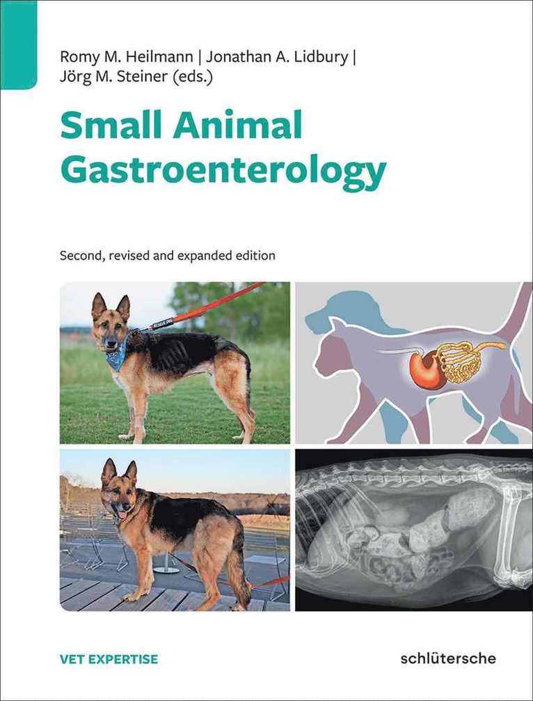


Du kanske också är intresserad av
Small Animal Gastroenterology
Romy M. Heilmann, Jonathan A. Lidbury, Jörg M. Steiner
Inbunden, 2024
2 299 kr
- Nyhet
Del 6