
Del i serien Diagnostic Imaging
Diagnostic Imaging: Breast
Inbunden, Engelska, 2019
4 499 kr
Beställningsvara. Skickas inom 5-8 vardagar. Fri frakt för medlemmar vid köp för minst 249 kr.
Covering the entire spectrum of this fast-changing field, Diagnostic Imaging: Breast, third edition, is an invaluable resource not only for radiologists, but for all health care professionals involved in the management of breast disease. From screening and diagnostic mammography and tomosynthesis, ultrasound, and MR to contrast-enhanced mammography and molecular imaging, Drs. Wendie Berg and Jessica Leung, along with their expert author team, provide carefully updated information in a concise, bulleted format. Thousands of high-quality illustrations highlight not only image acquisition and interpretation, but also screening guidelines, breast anatomy, genetic testing, image-guided procedures, determining the extent of disease and much more. This book provides essential, clinically-focused details for everyday breast imaging.
Produktinformation
- Utgivningsdatum2019-09-05
- Mått216 x 276 x 62 mm
- Vikt3 990 g
- FormatInbunden
- SpråkEngelska
- SerieDiagnostic Imaging
- Antal sidor1 350
- Upplaga3
- FörlagElsevier Health Sciences
- ISBN9780323548120
Tillhör följande kategorier
Hoppa över listan

Mer från samma serie
Diagnostic Imaging: Pediatrics
A. Carlson Merrow Jr., Michael R. Aquino, Luke L. Linscott, Bernadette L. Koch
Inbunden, 2022
4 529 kr
Diagnostic Imaging: Chest
Melissa L. Rosado-de-Christenson, Santiago Mart�nez-Jim�nez
Inbunden, 2021
4 699 kr
Diagnostic Imaging: Musculoskeletal Non-Traumatic Disease
Kirkland W. Davis, Donna G. Blankenbaker, Stephanie Bernard
Inbunden, 2022
4 679 kr
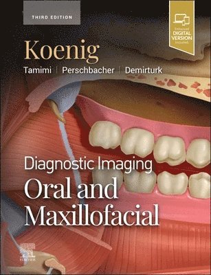
Diagnostic Imaging: Oral and Maxillofacial
Lisa J. Koenig, Dania Tamimi, Susanne E. Perschbacher, Husniye Demirturk
Inbunden, 2023
4 649 kr
Hoppa över listan

Du kanske också är intresserad av
Diagnostic Imaging: Musculoskeletal Trauma
Kambiz Motamedi, Donna G. Blankenbaker, Kirkland W. Davis
Inbunden, 2026
4 889 kr
Diagnostic Imaging: Pediatrics
A. Carlson Merrow Jr., Randy R. Richardson, Arthur B. Meyers, Michele Retrouvey, Lindsey Pelissier
Inbunden, 2027
4 379 kr
Diagnostic Imaging: Head and Neck
Bronwyn E. Hamilton, Bernadette L. Koch, Surjith Vattoth, Blair A. Winegar
Inbunden, 2025
4 659 kr
Diagnostic Imaging: Oral and Maxillofacial
Lisa J. Koenig, Dania Tamimi, Susanne E. Perschbacher, Husniye Demirturk
Inbunden, 2023
4 649 kr
Diagnostic Imaging: Pediatrics
A. Carlson Merrow Jr., Michael R. Aquino, Luke L. Linscott, Bernadette L. Koch
Inbunden, 2022
4 529 kr